Clinical Phenotyping Core
Core Mission
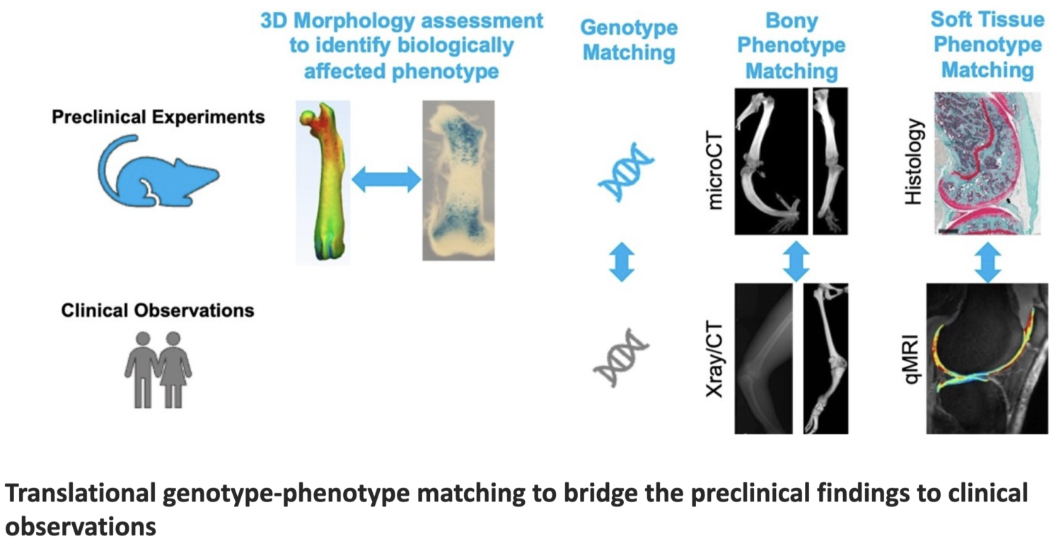
The Clinical Phenotyping Core offers a range of services (outlined below) focused on bridging the pre-clinical findings to clinical observations along with image-based assessments of joint morphology and soft tissue structures (e.g., tendons, ligaments and cartilage). The core also facilitates the use of largescale clinical databases to enable phenotype-genotype matching between pre-clinical and clinical observations (e.g., to see how observed phenotypes in a genetically modified animal models would compare to those seen in humans with the same genetic mutation).
Through these serveries, the core supports the CSR community with efforts related to studying the disease mechanism, imaging biomarker development and validation, and assessment of treatment outcomes for a range of skeletal disorders. Moreover, the core offers unique training opportunities through hands on training sessions and consulting.
Services
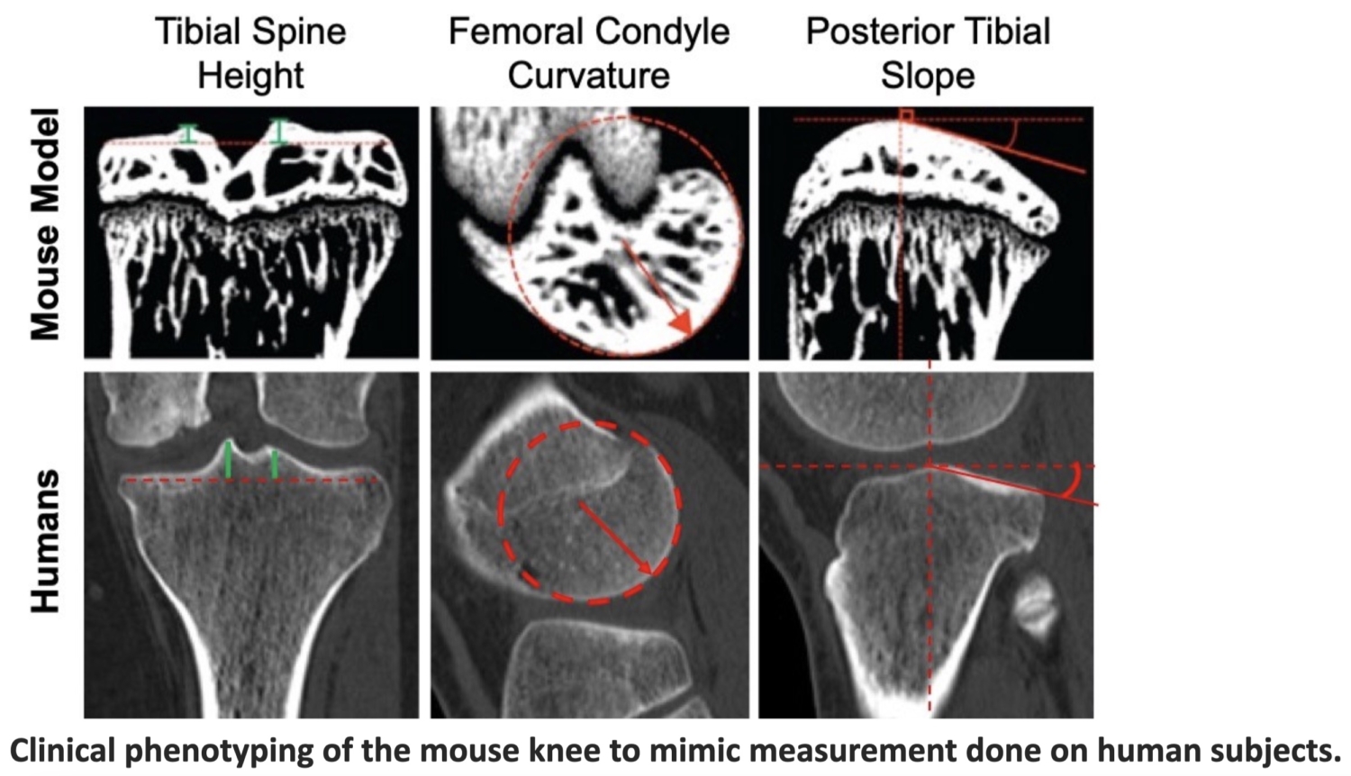
- 2D and 3D phenotyping of bony and soft tissue structures (e.g., bone shape, cartilage thickness) using clinically relevant measurement techniques to mimic clinical evaluations.
- Quantitative and semi-quantitative image analysis to assess tissue quality (e.g., cartilage and ligament) and remodeling from imaging data.
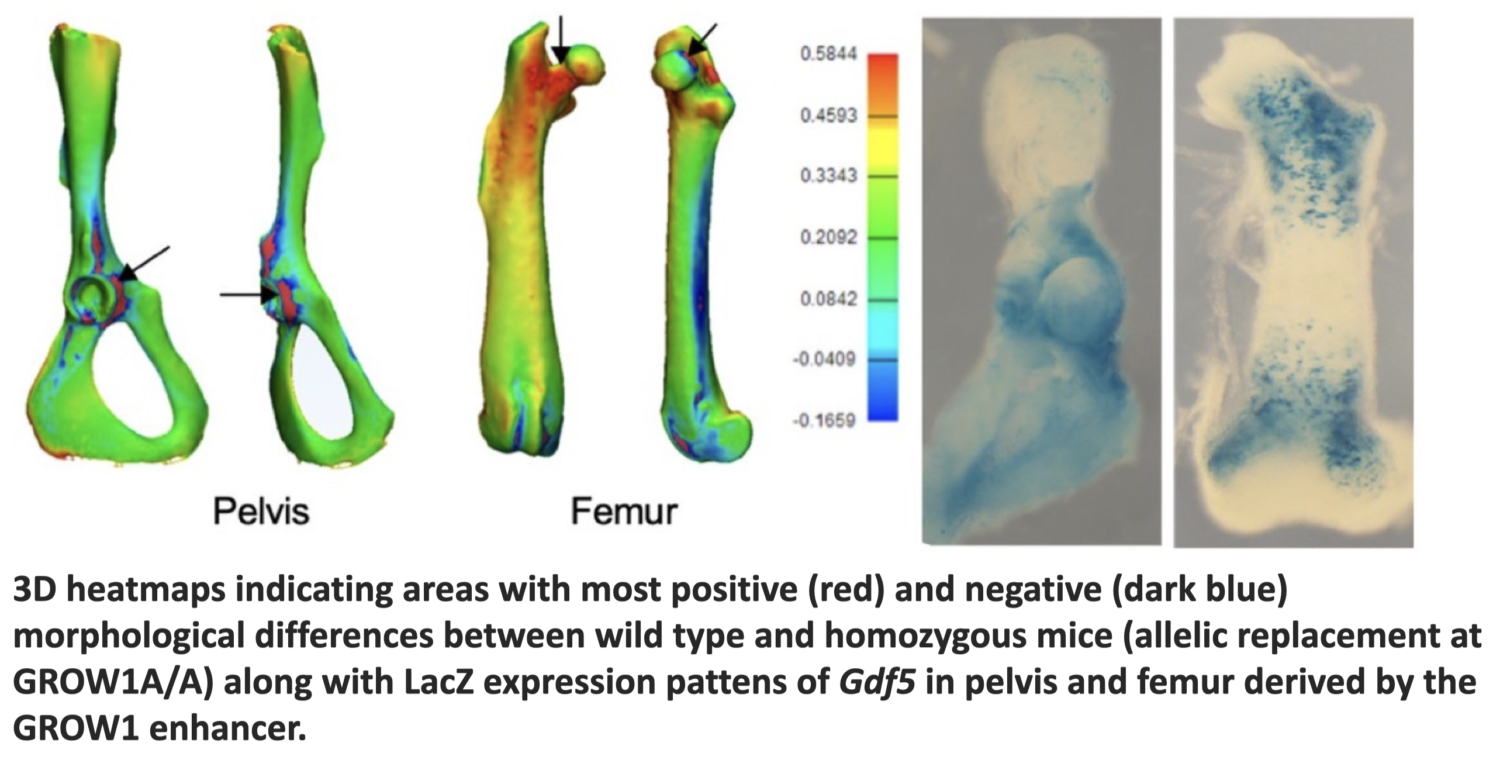
- Deep phenotyping (e.g., 3D mapping) to study complex morphologies.
- Imaging biomarker identification to predict the risk of injury or treatment outcomes.
- Genotype-phenotype matching of pre-clinical models (e.g., genetically modified mouse models) to clinical cohorts (e.g., patients with certain musculoskeletal disorders) to help better interpret and validate the preclinical observations.
- Evaluation of the normal and pathologic development of joint and musculoskeletal tissue structures along with their associated factors (e.g., genetics, injuries) by leveraging the unique clinical data at Boston Children’s Hospital*
- Assistance with processing the medical images of publicly available largescale clinical cohorts (e.g., OAI, UKBiobank)*
- Assistant with study design, outcome measure selection, imaging protocol development and analytical plans.
- Training on phenotyping and image processing of both preclinical and clinical imaging data.
* Following IRB and other relevant institutional approvals.
Equipment and Resources
- Image Processing and AI Workstations
- Image Processing Software Packages
- Small Animal 7T MRI System (Small Animal Imaging Lab)
- Access to a unique large dataset of clinical, imaging, and genetics and genomics data from patients (children and adults) with a range of musculoskeletal disorders
For customized project costs please contact Dr. Kiapour (ata.kiapour@childrens.harvard.edu)


