HR-pQCT Core
Core Mission
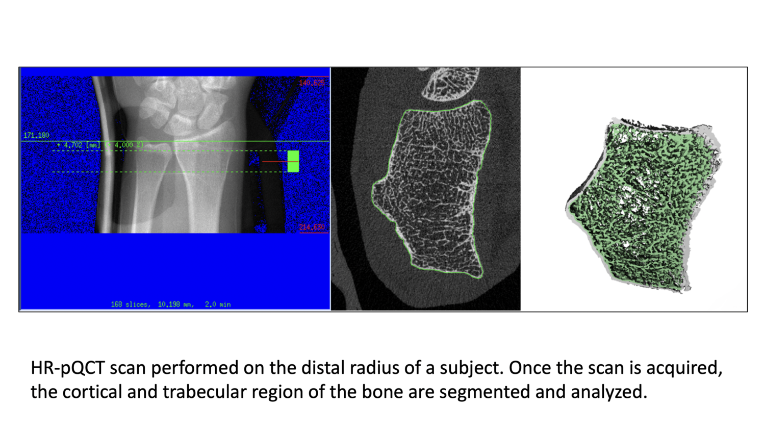
The High Resolution Peripheral Quantitative Computed Tomography (HR-pQCT) Core Facility provides clinical investigators with access to high resolution bone imaging. HR-pQCT allows for the measurement of bone density and microarchitecture of cortical and trabecular bone in the distal radius and tibia. Additionally, the HR-pQCT Core Facility offers Finite Element Analysis to estimate bone strength.
A scanning appointment is ~30 minutes and involves less radiation exposure than traditional bone densitometry. With applications for both cross-sectional and longitudinal research, investigators have employed our facilities and expertise to study a variety of bone-related diseases with participants ranging in age from children to older adults.
Services
- Assessment of bone density and microarchitecture for total, cortical and trabecular bone compartments of the distal radius and tibia.
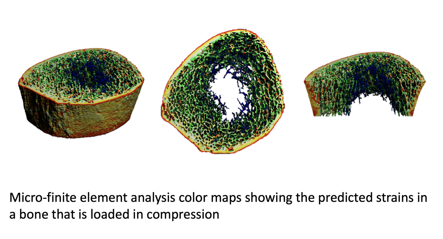
- Micro-Finite Element Analysis (µFEA): Estimates of bone stiffness and strength via micro-finite element analysis of the HR-pQCT images
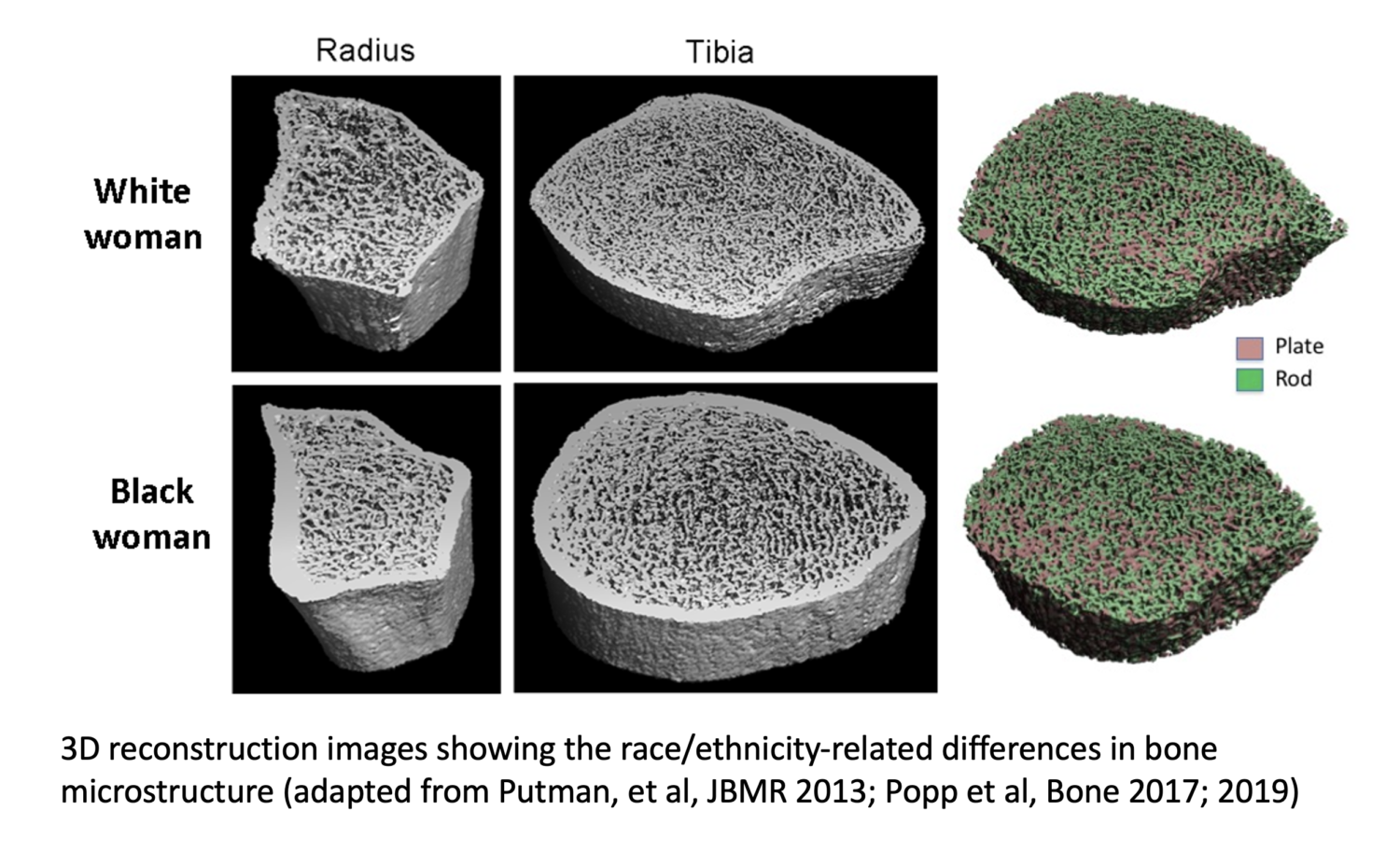
- Individual trabecular segmentation (ITS) analyses provided to characterize the rod- and plate-like nature of trabecular bone
- Two and 3D registration available for longitudinal studies
- Normative data (mean values and standard deviations) for healthy 20 to 30 year old men and women are available to provide reference values. Short-term reproducibility data are also available.
- Images for presentations: The HR-pQCT Core will work with investigators to generate high-quality images illustrating suitable for publications and presentations.
Equipment and Resources
Our facility has a Scanco XtremeCT II HR-pQCT scanner. This is the newest generation of Scanco’s HR-pQCT scanners and allows for faster and higher resolution scanning than the previous generation. This scanner is coupled with a computational cluster that provides high-speed reconstruction and analysis of scans.


